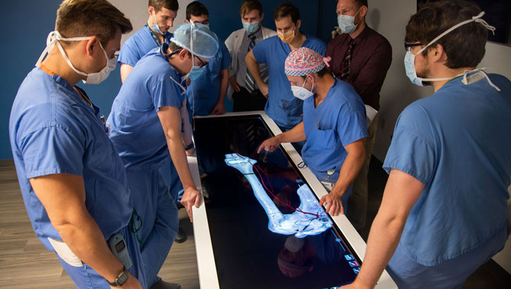
3D Analysis

3D Analysis is the most advanced orthodontic analysis tool on the market. Utilizing unique 3D tracing methods, users can create more accurate and consistent tracings directly from the cone-beam CT scan. Many schools and researchers choose Anatomage’s 3D Analysis module as their main research and publication tool. Surgeons can quickly create surgical cuts and patient soft tissue simulations, making it a very powerful and accurate consultation tool. 3D Analysis can also be used for tracing standard 2D analyses, eliminating the need for reconstructing and tracing on ambiguous 2D X-ray images.

Comprehensive 3D Analysis

Visualize your cephalometric analyses in 3D space with our extensive and comprehensive landmark and measurement library or modify and create your own analysis.


Photo Face Wrap

Superimpose a traditional clinical 2D photograph or a 3D facial image onto your patient’s scan to visualize soft tissue features and create the ultimate virtual patient.

Impress Your Patients

Simulate soft-tissue movements with real-time orthognathic surgical cuts. Rotate and translate with accurate re-positioning tools to enhance your consultation.

Assisted Landmark Identification

Anatomical landmarks have always been difficult to identify on flat cephalometric scans. To solve this problem, Anatomage created an innovative and simple way to perform cephalometric tracings in 3D that will automatically identify many specific points. Our comprehensive landmark and angle measurement library will allow you to accurately position points and easily define profiles on a 3D scan.

  • Simplify Cephalometric Tracings in 3D
  • Mathematically Accurate Landmark Identification
  • Intuitive Interface for You and Your Staff

Create Your Custom 3D Analysis

As an experienced clinician, you already understand your patient’s skeletal proportions, asymmetries, and soft-tissue profile. However, formalizing your understanding into concrete data across multiple patients can be very difficult without the help of powerful software. With the 3D Analysis add-on module, we have addressed this problem and made it easy to establish your own measurements and build your norm data statistics over time. Whether you’re creating a research project or building upon existing measurements, 3D Analysis will assist you in effectively creating a custom analysis.

  • Customize Your 3D Measurements
  • Build & Analyze Your Norm Data
  • Easily Export Your Results

Fully Compatible with traditional 2D Analysis

Because the precedence for 2D analysis and the volume of existing data for 2D analysis are still important, the 3D Analysis module also provides the full traditional 2D analysis as well. The software comes set up with the most commonly used analyses and lets you easily create your own or modify existing ones. Here is a partial list of included analyses and data sets:

  • Steiner
  • ABO
  • Alexander
  • Downs
  • Tweed
  • UOP
  • Ricketts
  • Bjork
  • Iowa
  • McNamara
  • McLaughlin
  • Sassouni

Automatic 3D Surgical Cuts

Once the anatomical landmarks have been identified, you can easily visualize orthognathic surgery simulations in 3D. There is no need to tediously construct an iso-surface and manually define surgical cuts, as this is calculated automatically based on the geometry of your tracings.  Accurately reposition maxillary, mandibular, and chin cuts to analyze how simulated movements will affect landmark position, profile lines, and the superimposed patient image.

  • Automatic Surgical Cuts Allows for Surgical Simulations
  • Fast and Immediately Ready for Surgical Analysis
  • Accurate Re-positioning

Photo Wrapping & Soft Tissue Simulations

The 3D Analysis module can create 3D photos using a CBCT scan and a standard digital photo. The constructed 3D surface is accurate since it is derived from the CBCT scan and perfectly aligned to the underlying hard tissue. This module allows patient 3D soft tissue simulations with surgical analysis. When a surgical analysis is performed the soft tissue is updated in real time. The movement could be in any 3D orientation and the soft tissue change will be smooth and realistic.

  • Create 3D Photos
  • Simulate Real time Patient Skin Movement
  • Powerful Case Presentation Tool
Want to Schedule a Meeting?