
The Anatomage Table gives students at Misericordia University the tools to perform virtual dissections on a human cadaver, just as they would in a regular medical classroom. The Table provides future healthcare professionals a sense of autonomy as they utilize the 3D virtual technology for independent studying.
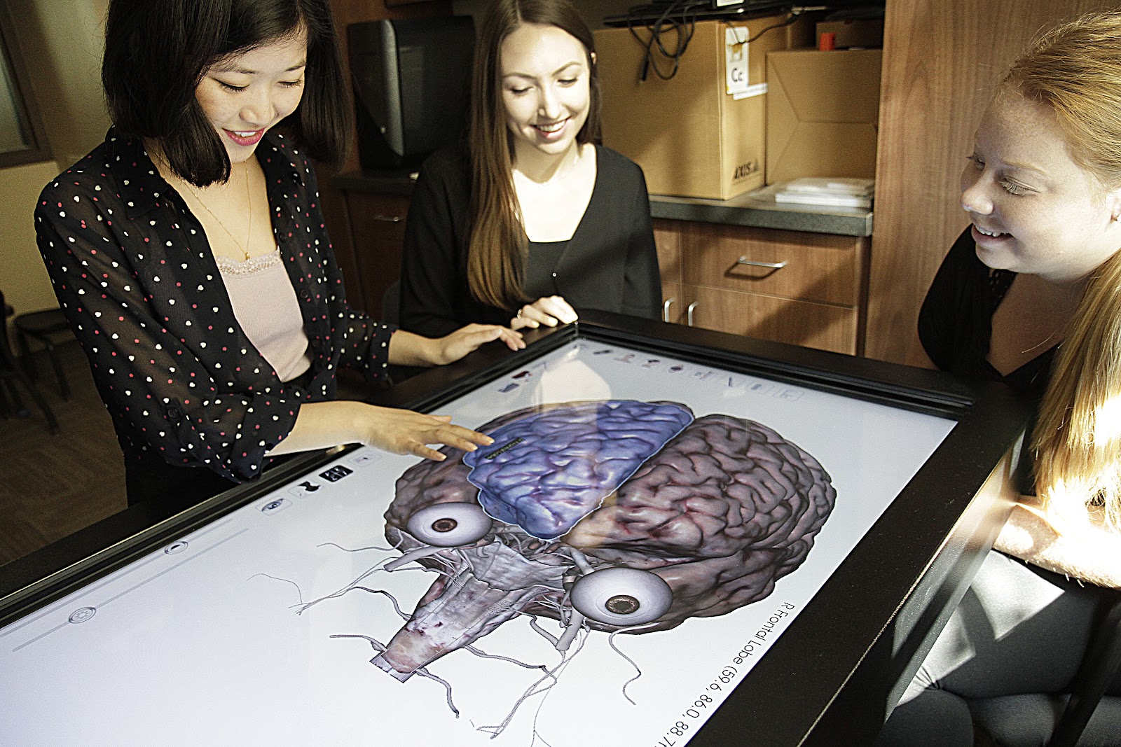
Jillian Scanlon and Annette Ritzko, speech-language pathology students, from Misericordia University are known at the university as “master users” of the Anatomage Table. Not only do they teach fellow students how to use the Table but they also teach university instructors and sometimes create classroom materials for them.
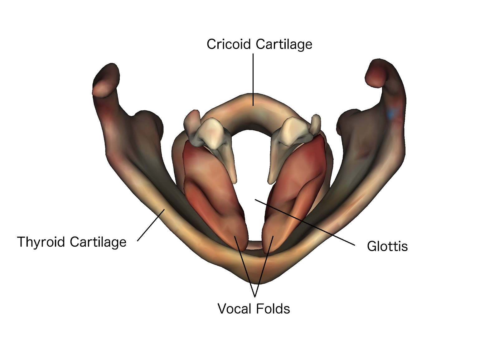
Using the head and neck model of the Anatomage Table, the segmented body feature allows university students to study in-depth anatomy surrounding speech and swallowing disorders. Learning how a stroke can lead to someone being diagnosed with aphasia may be difficult to comprehend if using a traditional textbook and 2D images. With the Anatomage Table, speech-language pathology students can visualize the spatial relationships of anatomical structures such as the head, neck, and lung. Isolating anatomical structures can provide university students with the capabilities to shift their concentration to body segments they are studying.
Dr. Glen Tellis is a professor and chairman of the Speech-Language Department at Misericordia University. Encouraging his students to use technology to break out of the conventional way of learning, Dr. Tellis utilizes the Anatomage Table to warrant his students to teach each other comprehensive anatomy. Having an unorthodox approach to teaching in-depth anatomy has empowered his students to leverage the Anatomage Table as a tool for their anatomy education.
At Misericordia University, fifth-year graduate student at the speech-language pathology department, Annette Ritzko, uses the Anatomage Table to research the anatomy of the human brain and the esophagus to better comprehend speech and swallowing disorders. Focusing on the larynx and pharynx to show in-depth anatomy, Annette learns more than just high level anatomical bodily structures. Being a “ master user” of the Anatomage Tale, Annette created a user manual for other students to learn the function of the Table to further study highly detailed anatomy. Annette prepares students by giving them the manual that outlines the steps of how to use the several features and capabilities of the Table. This is then followed up by an hour-long hands-on experience with the Anatomage Table. Annette teaches her classmates how to leverage Anatomage’s technology for independent studying and teaching. The virtual dissection tools in the Anatomage Table encourages students to become more engaged in their learning.
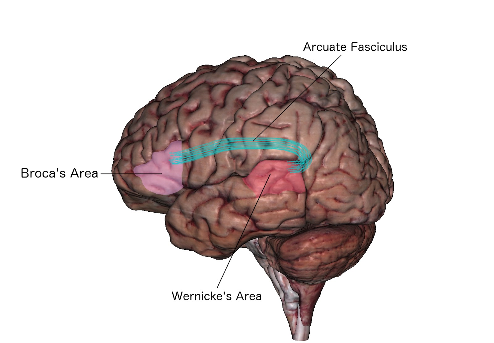
Pictured above is an image from the Anatomage Table that was created by one of the speech-language pathology students. The following is an example of the types of learning material that is created using the capture tool. 3D models of regional anatomy enable students to easily personalize study material and share with their classmates.
Still skeptical? Ask Jillian Scanlon, who will complete her last year as an undergraduate student in the speech-language department. Jillian was recruited by both Annette and Dr. Tellis to be a part of their research clinic. Jillian states “anatomy and physiology is the foundation of what we do [as speech-language pathologists]. The Anatomage Table helps me better learn and remember anatomy and physiology. It’s so engaging and that helps!” During the first month and a half of her sophomore year, Jillian used traditional learning from a textbook to study anatomy. She would also use google search engine to see 2D images of anatomical structures, such as the cranial nerve, and hoped it was enough for her to study. As she took anatomy and physiology exams, she passed with an average of 87%. Soon after using the Anatomage Table to study, she began getting 99% and 100% on all of her exams. Nearing a perfect score in all of her tests, she credits the Anatomage Table for helping her comprehend highly detailed anatomical structures in the human body.
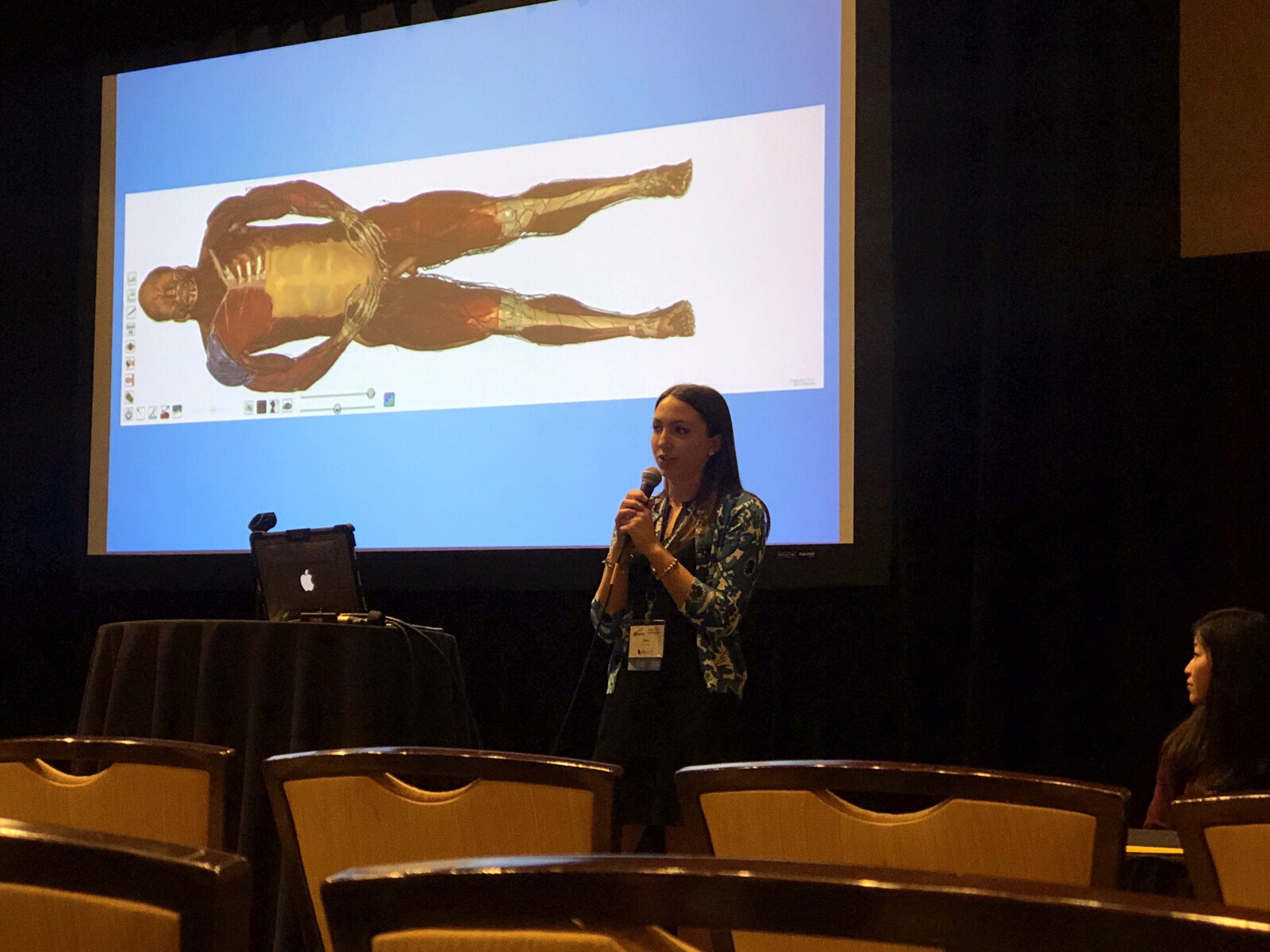
Since using the Anatomage Table, Jillian has joined Annette in presenting at conferences, such as the Pennsylvania Speech-Language-Hearing Association. At the conference, Jillian and Annette discussed how education technology tools like the Anatomage Table empowers students to have a sense of ownership when it comes to their learning capabilities. Anatomage is proud to support future healthcare professionals by creating technology that surpasses traditional learning and teaching methods.